
Image Credit: 3DPrint.com
Last month, a relative underwent what was expected to be a routine ablation procedure: 9 1/2 hours and 3 a-fib episodes later, the surgery finally finished. Despite CT scans, X rays and EKGs, the surgeons encountered "structural issues" that complicated the operation. I thought afterward if they had had a 3D print of his heart, they might have anticipated and planned contingencies based on what they saw. Apparently I'm not alone in this believe...
A masters student from Drexel, Jason Kirk, released a study, "3D Printed Cardiac Imaging Data," that suggests that patients and surgeons benefit from reviewing patient-specific 3D printed replicas of their organs prior to consenting to surgery. Feedback given to the researcher indicates that a majority of surgeons find 3D models more effective than 2D illustrations in sharing information and facilitating discussions. According to Kirk, “Cardiac anatomy replicas can be used to facilitate Doctor/Patient communication and supplement contemporary visualization techniques by providing accurate three dimensional data which offers additional haptic and spatial feedback specific to the patient’s anatomy and pathology.”
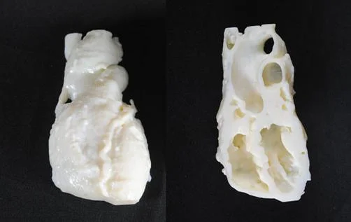
But how is the replica made? CT scans and MiMiC software are used to create custom 3D prints that can include cut aways to show the internal structure of the organs. And the practice is becoming more popular: RapidMade recently created lung models for a research center interested in using them for patient education.